Glaucoma : Guide to Diagnosis, Treatment, and Prevention
675
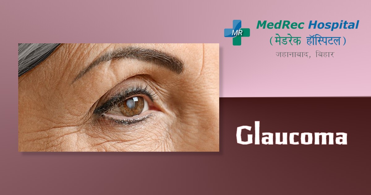
The optic nerve is harmed by glaucoma, a group of eye diseases. The optic nerve, which sends visual information from the eye to the brain, is crucial for seeing clearly. Damage to the optic nerve is usually linked to high eye pressure. However, glaucoma can develop with normal eye pressure as well.
Even while it may strike anybody, older persons are more likely to develop glaucoma. It is one of the major causes of blindness in those over 60.
Numerous glaucoma types show no symptoms at all. You might not notice a change in vision until the problem is advanced since the effect is so subtle.
You should get regular eye exams and have your eyes measured.
Early glaucoma diagnosis allows for possible prevention or slowing of visual loss. For the remainder of their life, glaucoma patients will require medication or monitoring.
Causes
When the optic nerve is harmed, glaucoma results. When this nerve gradually deteriorates, blind spots start to emerge in your vision. This nerve injury is typically correlated with elevated ocular pressure for reasons that doctors are unsure of.
Elevated intraocular pressure results from a buildup of fluid that moves around the eye's interior. Another term for this material is aqueous humour. It usually leaves through a tissue where the iris and cornea meet. This tissue is also known as the trabecular meshwork. As it allows light to enter the eye, the cornea is crucial to vision. Eye pressure may rise when the eye produces too much fluid or when the drainage system is not functioning properly.
Angle-opening glaucoma
This is the most prevalent kind of glaucoma. The drainage angle between the iris and cornea is still open. However, several components of the drainage system aren't working properly. This might lead to a gradual rise in ocular pressure.
Glaucoma with angle closure
This kind of glaucoma manifests as iris enlargement. The drooping iris partly or completely blocks the drainage angle. The pressure inside the eye increases as a result, and fluid cannot pass through it. Angle-closure glaucoma may progress rapidly or gradually.
Glaucoma with normal-tension
Nobody is exactly sure why the optic nerve is hurt when the eye pressure is normal. It is possible that the optic nerve's blood supply will be less or more sensitive. The accumulation of fatty deposits in the arteries may be the source of this constrained blood flow.
Children's glaucoma
Glaucoma can either be present from birth or appear in the first few years of life in a kid. Obstructed drainage, trauma, or an underlying medical condition can all harm the optic nerve.
Pigmented glaucoma
Pigmentary glaucoma is characterized by small pigment granules that detach from the iris and impede or slow fluid expulsion from the eye. The pigment granules might occasionally be stirred up by activities like jogging. As a result, pigment granules are placed on the tissue at the iris and corneal junction. The granule deposition causes an increase in pressure.
As a rule, glaucoma runs in families. Scientists have discovered genes that are connected to damaged optic nerves and elevated eye pressure in some individuals.
How to check if you have Glaucoma?
Acute angle-closure glaucoma may be the cause of the abrupt onset of symptoms. Two symptoms include a severe headache and eye pain. Go to the emergency hospital or make a quick call to an ophthalmologist's office.
Risk Factors
Glaucoma can affect vision even before any symptoms manifest. Consider these risk variables, then:
- High internal eye pressure, often known as intraocular pressure,
- Older than 55
- Familial glaucoma history
- Several medical disorders, including high blood pressure, diabetes, migraines, and sickle cell anaemia
- Centres of corneas that are thin
- Either extreme near- or farsightedness
- Eye damage or specific eye surgeries
- Corticosteroid drug usage over a long period of time, especially eye drops
- Some people are more prone to developing angle-closure glaucoma because they have limited drainage angles.
Symptoms
Depending on the kind and stage of your ailment, your glaucoma symptoms will vary.
Open Angle Glaucoma
- Early stages with no symptoms
- Patchy blind patches appear in your side vision gradually.
- Peripheral vision is another name for side vision.
- Difficulties seeing details in your centre vision as the disease progresses
Glaucoma with acute angle-closure
- Terrible headache
- Terrible eye discomfort
- Nausea or diarrhoea
- Distorted vision
- Rings of colour or halo effects surrounding lights
- A reddened eye
- Glaucoma with normal-tension
- Early stages with no symptoms
- Vision gradually becomes hazy
- Later, side vision may be lost
Children's glaucoma
- A hazy or drab eye (infants)
- Higher blink rate (infants)
- Tears (infants)
- Distorted vision
- Increasing nearsightedness
- Headache
Prevention
By taking these steps, glaucoma may be identified and treated earlier. That might decrease the deterioration of vision or assist to avoid it.
Regularly check your eyes. Regular, thorough eye exams can aid in identifying glaucoma in its early stages before serious damage takes place.
You will require more regular screening if you are at risk for glaucoma. Ask your doctor to suggest the appropriate screening schedule. with you.
Know the history of eye disease in your family. As a rule, glaucoma runs in families. If your risk is higher, you might require more regular screening.
Invest in eye protection. Glaucoma may result from severe eye damage. When using power tools or engaging in sports, use eye protection.
Frequent use of eye drops is prescribed. The likelihood that elevated eye pressure may develop into glaucoma can be considerably decreased by using glaucoma eye drops. Even if you are symptom-free, use eye drops as directed by your doctor.
Treatments
Glaucoma can not be treated, thus the damage is irreversible. However, if you discover the condition in its early stages, medication and routine exams can help delay or prevent vision loss.
In order to treat glaucoma, intraocular pressure is reduced. Prescription eye drops, oral medications, laser therapy, surgery, or a combination of methods are all available as treatment options.
Eyedrops
Prescription eye drops are frequently the first step in glaucoma therapy. Some people may lower eye pressure by enhancing the eye's ability to discharge fluid. Others lessen the fluid production in your eye. Depending on how low your eye pressure needs to be, you could need more than one eye drop.
Among the prescription eye drops are:
Prostaglandins. These enhance the fluid in your eye's outflow, which aids in lowering ocular pressure. Medicines: Latanoprost (Xalatan), travoprost (Travatan Z), tafluprost (Zioptan), bimatoprost (Lumigan), and latanoprostene bunod are among the medications in this category (Vyzulta).
Mild eye reddening and stinging, iris darkening, eyelash or eyelid skin pigment darkening, and impaired vision are all potential adverse effects. One dose per day is recommended for this kind of medication.
Beta Blockers. These lessen the amount of fluid that is produced in your eyes, which helps to alleviate ocular pressure. Timolol (Betimol, Istalol, Timoptic) and betaxolol are two examples (Betoptic S).
Breathing issues, a slower heartbeat, reduced blood pressure, impotence, and weariness are all potential adverse effects. Depending on your condition, this class of medication may be given for once- or twice-daily usage.
Agonists of the alpha receptor. These lessen the amount of fluid produced that circulates within your eye. Additionally, they cause your eye's moisture to drain more quickly. Examples include brimonidine and apraclonidine (Iopidine) (Alphagan P, Qoliana).
Uneven heartbeat, elevated blood pressure, exhaustion, red, itchy, or swollen eyes, and dry lips are a few potential adverse effects. Although occasionally three times a day, this type of medication is often given for twice daily usage.
Blockers of carbonic anhydrase. These drugs lessen the amount of fluid that is produced in your eyes. Dorzolamide and brinzolamide are two examples (Azopt). A metallic taste, frequent urination, and tingling in the fingers and toes are a few potential adverse effects. Although occasionally three times a day, this type of medication is often given for twice daily usage.
Inhibitor of rho kinase. This medication reduces eye pressure by inhibiting the rho kinase enzymes in charge of fluid expansion. It comes in the form of netarsudil (Rhopressa), and a daily dose is recommended. Red eyes and pain in the eyes are potential adverse effects.
Cholinergic or miotic substances. These cause your eye's fluid discharge to rise. Pilocarpine is an illustration (Isopto Carpine). Headache, eye pain, smaller pupils, potentially impaired or dim vision, and nearsightedness are some of the side effects. The typical dosage for this kind of medication is up to four times per day. These medications are no longer prescribed very frequently due to possible adverse effects and the requirement for repeated daily usage.
You can encounter certain adverse effects unrelated to your eyes since part of the eye drop medication is absorbed into your system. After applying the drops, shut your eyes for 1 to 2 minutes to reduce this absorption. To seal the tear duct for a minute or two, you can gently lightly press on the corner of your eyes next to your nose. Remove any droplets that are not needed from your eyelid.
You might need to utilise artificial tears or several eye drops that have been given to you. Be careful to give yourself at least five minutes in between each drop application.
Drugs administered orally
Your eye pressure may not decrease to the appropriate level with only eye drops. So, your eye doctor could also recommend using oral medication. Usually a carbonic anhydrase inhibitor. Frequent urination, tingling in the fingers and toes, sadness, stomach trouble, and renal problems are examples of potential adverse effects.
Operations and other treatments
Laser therapy and surgery are other therapeutic possibilities. The following methods might aid in dripping fluid from the eye and bringing down eye pressure:
Laser treatment. You have the option of laser trabeculoplasty if you cannot tolerate eye drops. If medication has not halted the development of your condition, it may also be utilised. Before utilising eye drops, your eye doctor may possibly suggest laser surgery. It happens in the office of your eye doctor. The drainage of the tissue at the point where the iris and cornea meet is improved by your eye specialist using a little laser. Before the entire impact of this treatment is felt, it can take a few weeks to filter. This is a trabeculectomy, a
Surgery
The sclera, often known as the white of the eye, is opened by the eye surgeon. The procedure makes a second opening for fluid to exit the eye.
tubes for draining. In order to drain extra fluid and relieve eye pressure, the eye surgeon inserts a tiny tube into your eye during this surgery.
Glaucoma surgery with minimum disruption (MIGS). To reduce your eye pressure, your eye doctor could advise undergoing a MIGS surgery. Compared to trabeculectomy or the use of a drainage device, these treatments often need less immediate postoperative care and have fewer risks. They frequently accompany cataract surgery. Your eye doctor will explain which MIGS technique could be best for you out of the several ones that are now available.
Following your surgery, you must visit your eye doctor for further examinations. If your eye pressure ultimately starts to rise or if other changes take place in your eye, you could eventually need to undergo further treatments.
Acute angle-closure glaucoma treatment
Glaucoma with acute angle closure is a medical emergency. In order to lower the pressure in your eye if you are diagnosed with this illness, you will require immediate treatment. Typically, this calls for either medical intervention, laser surgery, or both.
A technique known as a laser peripheral iridotomy can be performed on you. The medical professional uses a laser to make a tiny hole in your iris. As a result, liquid can pass through the iris. This lowers ocular strain and aids in opening the drainage angle of the eye.
For further information please access the following resources:
Emergency : +91 89686 77907
Front Desk : +91 98018 79584
Page last reviewed: Mar 14, 2023
Next review due: Mar 14, 2025


.jpg)
.jpg)
.jpg)
.jpg)
.jpg)
.jpg)