


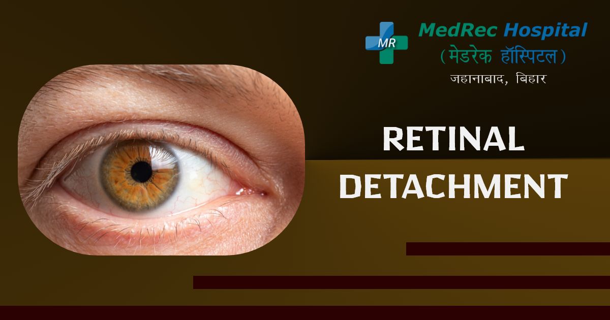
Retinal Detachment Prevention, Symptoms And Treatment
691
A thin layer of tissue (the retina) in the back of the eye slips away from its usual position in an emergency scenario known as retinal detachment.
The layer of blood vessels that feeds and oxygenates the eye is separated from the retinal cells by retinal detachment. The risk of irreversible vision loss in the afflicted eye increases the longer retinal detachment is left untreated.
Potential warning signs of retinal detachment include reduced vision, the abrupt appearance of floaters, and bright flashes. You can save your vision by calling an ophthalmologist (eye expert) straight away.
Causes
There are three forms of retinal detachments:
Rhegmatogenous. These are the retinal detachments that occur most often. A hole or tear in the retina that permits fluid to pass through and pool beneath the retina is what causes rhegmatogenous detachments. The outcome of this fluid buildup is that the retina starts to separate from the supporting structures. You lose eyesight because the retinal detachment sites experience a reduction in blood flow and functional decline.
The most frequent cause of rhegmatogenous separation is old age. As you age, the vitreous, a gel-like material that coats the inside of the eye, may lose volume or become more liquid. In most cases, the vitreous separates from the retina's surface without any issues.
Posterior vitreous detachment (PVD). A rip is one issue with this separation.
A retinal tear may result from the vitreous pulling on the retina as it separates or peels off. If the tear is not repaired, the liquid vitreous may leak through it and into the area behind the retina, causing the retina to detach.
Tractional. As scar tissue develops on the retina's surface, the retina might detach from the back of the eye and peel away. Those with poorly managed diabetes or other illnesses frequently have tractional separation.
Exudative. Instead of leaving holes or tears in the retina, this kind of detachment causes fluid to build up behind the retina. Tumors, inflammatory conditions, age-related macular degeneration, ocular traumas, or age-related macular degeneration can all cause exudative disassociation.
How to check if you have Retinal Detachment?
If you see any of the warning signs or symptoms of retinal detachment, get medical help right once. A medical emergency called retinal detachment can result in irreversible eyesight loss.
Risk Factors
The risk of retinal detachment is increased by the following factors:
- Retinal detachment is more frequent in adults over 50 due to aging.
- Prior retinal detachment in one eye; retinal detachment in the family
- Extreme nearsightedness (myopia)
- Earlier eye procedures, such as the removal of a cataract
- Previous serious eye injury
- Prior eye conditions such as retinoschisis, uveitis, or thinning of the peripheral retina (lattice degeneration)
Symptoms
The actual retinal detachment causes no discomfort. Yet before it happens or has progressed, there are virtually always warning indicators, such as:
- The unexpected emergence of numerous floaters, which are little specks that seem to be moving through your field of vision
- Flashes of light in either one or both eyes (photopsia)
- Blurry vision
- Gradually deteriorating side (peripheral) eyesight
- Curtain-like darkness blocked your field of vision.
Treatments
A retinal tear, perforation, or detachment is usually invariably repaired surgically. There are several methods accessible. Discuss the advantages and disadvantages of your treatment choices with your ophthalmologist. Together, you may decide which surgery or set of procedures is ideal for you.
Tears in the retina
Your eye surgeon may advise one of the following procedures to stop retinal detachment and maintain vision if a retinal tear or hole has not yet developed into a detachment.
Laser treatment (photocoagulation). Via the pupil, the surgeon shines a laser into the patient's eye. The laser burns the area around the retinal tear, causing scarring that typically "holds" the retina to the underlying tissue.
Freezing (cryopexy). The surgeon uses a freezing probe to numb your eye after administering a local anesthetic. The area of the eye's exterior right above the tear. The scar left behind by the freezing serves to anchor the retina to the eye wall.
Both of these operations are performed as outpatients. After your treatment, you will probably be told to refrain from eye-jarring activities like running for a few weeks.
Retinal detachment
If your retina has detached, surgery to reattach it is required, ideally soon after a diagnosis. Many criteria, including the degree of the separation, will determine the kind of surgery your surgeon advises.
Injecting gas or air into your eye. A bubble of air or gas is injected into the eye during this treatment, known as pneumatic retinopexy.
The eye's central region (the vitreous cavity). If placed correctly, the bubble presses the retinal region that contains the hole or holes against the eye's wall, halting the passage of fluid into the space behind the retina. During the surgery, your doctor will also employ cryopexy to heal the retinal break.
The retina can then cling to the eye's wall when the fluid that had accumulated beneath it is naturally removed. To maintain the bubble in the correct position, you might have to hold your head in a particular posture for as long as several days. The bubble will ultimately self-resorb.
Your eye's surface is indented. Surgeons perform a surgery known as scleral buckling. Over the damaged region, stitch (suture) a piece of silicone material to the sclera of your eye. The vitreous pulling on the retina is lessened with this technique, which dimples the eye's wall.
Your surgeon may make a scleral buckle that belts your whole eye if you have several tears, holes, or a significant separation. The buckle is positioned such that it does not obstruct your eyesight, and it typically stays in place for good.
Draining and replenishing the fluid in the eye. The surgeon removes the vitreous along with any tissue that is pulling on the retina during this treatment, which is referred to as a vitrectomy. The vitreous is then injected with air, gas, or silicone oil.
The vitreous area will eventually be filled with bodily fluid when the air, gas, or liquid has been absorbed. If silicone oil was used, it could need to be surgically removed months later.
Scleral buckling surgery and vitrectomy may be coupled.
It could take many months for your vision to go better after surgery. For effective therapy, you could require a second operation. Some people never fully regain their vision.
For further information please access the following resources:
Emergency : +91 89686 77907
Front Desk : +91 98018 79584
Page last reviewed: Apr 7, 2023
Next review due: Apr 7, 2025
.jpg)
.jpg)
.jpg)
.jpg)
.jpg)
.jpg)